Humeral Fracture Fixation/Knowledge Review/Post-Operative Management
This module allows medical officers and surgeons who are not orthopedic specialists to become confident and competent in irrigation and debridement, powered and manual drilling, positioning and correctly inserting Schanz screws, and constructing the rod-to-rod modular frame as part of external fixation procedures for open humeral shaft fractures performed in regions without specialist coverage. To maximize patient safety, this module teaches learners to use a powered drill to insert self-drilling Schanz screws through the near cortex and then manually advance Schanz screws into the far cortex to avoid plunging.
Learning Objectives
By the end of this module, learners will be able to:
- Conduct a history and physical examination of a patient following modular external fixation of an open humeral shaft fracture.
- Review the post-operative anteroposterior and lateral view radiographs of a patient following modular external fixation of an open humeral shaft fracture.
- Provide post-operative care for a patient following modular external fixation of an open humeral shaft fracture.
- Know the indications for adjusting the modular external fixator for a patient with an open, simple humeral shaft fracture.
- Know the indications for referral of a patient with an open humeral shaft fracture to a tertiary center for specialist care.
- Know how to apply a U-shaped cast for a humeral shaft fracture.
Postoperative Clinical Assessment
It's highly recommended to print off this module, bring it on ward rounds and clinic visits, have it available when reviewing the patient's X-rays, and keep it filed in the patient's chart to use as a checklist during the post-operative and follow-up management of a patient with an open humeral shaft fracture.
Physical Examination
Neurovascular Exam
The incidence of radial nerve injury is as high as 60% in open humeral fractures..[1]
Vascular Exam
Compare both sides when evaluating radial artery pulses.
- Palpable versus Not Palpable
- Symmetric versus Asymmetric
If the radial artery pulse is not palpable, check capillary refill.
If the radial artery pulse is not palpable, check for signs of acute compartment syndrome.
Sensory Testing
To test the posterior cutaneous nerve of the radial nerve (C6), perform light touch sensation testing on the posterior forearm and compare it to the other side.[2]
- Intact versus Not Intact
- Symmetric versus Asymmetric
To test the superficial branch of the radial nerve (C6-C8), perform light touch sensation testing on the thumb, index, and middle finger, or radial half of the ring finger, and compare it to the other side.[2]
- Intact versus Not Intact
- Symmetric versus Asymmetric
Motor Testing
Ask patient to perform arm abduction for motor testing of the deltoid muscle. Be sure to compare both sides.
- Able versus Unable
- Symmetric versus Asymmetric
Ask patient to perform elbow extension for motor testing of the triceps muscles. Be sure to compare both sides.
- Able versus Unable
- Symmetric versus Asymmetric
Ask patient to perform elbow flexion for motor testing of the biceps muscles. Be sure to compare both sides.
- Able versus Unable
- Symmetric versus Asymmetric
Ask patient to perform wrist extension for motor testing of the wrist extensors. Be sure to compare both sides.
- Able versus Unable
- Symmetric versus Asymmetric
Deep Tendon Reflexes
Place your hand under the biceps tendon and strike the hammer on the thumb. Observe for contraction of the biceps muscle. Be sure to compare both sides.
Reflex Grading:
- 0 Absent
- 1+ Diminished
- 2+ Normal
- 3+ Increased
- 4+ Hyperactive
- 5+ Sustained clonus
- Symmetric versus Asymmetric
Other Testing
The acceptable reduction parameters for open humeral shaft fractures are:
- < 15° malrotation (at 0° of rotation the patient's palm is facing straight up towards the ceiling when the patient is supine and the forearm is supinated)
- < 3 cm limb shortening (cannot be measured intraoperatively)
Rotation
- < 15° malrotation (at 0° of rotation the patient's palm is facing straight up towards the ceiling when the patient is supine and the forearm is supinated)
- > 15° malrotation
Limb Length Discrepancy
- < 3 cm limb shortening
- > 3 cm limb shortening
If the reduced humeral shaft fracture is not within acceptable parameters, the patient must return to the operating room for re-adjustment of fracture fragments.
Acute Compartment Syndrome
Evaluate for symptoms of acute compartment syndrome.[3]
- Pain disproportionate to injury and intensified with passive stretch (i.e., flexion and extension of the toes)
- Pallor
- Paresthesias
- Paralysis
- Pulselessness
- Compartment pressure greater than 30-40 mmHg in an unconscious or paralyzed patient
Acute compartment syndrome is a surgical emergency and must be treated with a fasciotomy. If the patient has signs of acute compartment syndrome, the patient should be referred to a tertiary center for specialist care.
Skin Tenting
All pin sites will be inspected for skin tenting.
- Proximal Fragment
- Schanz Screw #1 (Far Pin)
- Schanz Screw #2 (Near Pin)
- Distal Fragment
- Schanz Screw #3 (Near Pin)
- Schanz Screw #4 (Far Pin)
If skin tenting is present, the stab incision should be widened to release any soft tissue tension around the pin site to reduce the risk of inflammation and pin infection.[4]
Proper Placement of Far Pin in Distal Fragment
Check whether the Schanz Screw #4 (Far Pin) in the distal fragment was placed at least 2 fingers’ breadth proximal to the lateral epicondyle.
- Distal Fragment Far Pin Placed At Least 2 Fingers' Breadth Proximal to Lateral Epicondyle
- Distal Fragment Far Pin Not Placed At Least 2 Fingers' Breadth Proximal to Lateral Epicondyle
Always review the post-operative X-ray to check whether the pin has entered the elbow joint.
Pins Inserted In Safe Zones of the Humerus
Avoiding Axillary Nerve Injury
- Proximal Fragment Far Pin Inserted At Least 7 cm Below the Acromion
- Proximal Fragment Far Pin Not Inserted At Least 7 cm Below the Acromion
Avoiding Radial Nerve Injury
- Proximal Fragment Near and Far Pins Inserted Anterolaterally
- Proximal Fragment Near and Far Pins Not Inserted Anterolaterally
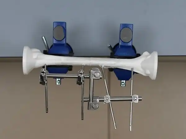
Frame Loosening
Check that all the clamps are securely tightened. Use the 11 mm spanner with T handle to tighten clamps at the bedside, if required.
- Proximal Fragment Rod
- Schanz Screw #1 (Far Pin) Clamp
- Schanz Screw #2 (Near Pin) Clamp
- Distal Fragment Rod
- Schanz Screw #3 (Near Pin) Clamp
- Schanz Screw #4 (Far Pin) Clamp
- Connecting Rod
- Proximal Rod-to-Rod Clamp
- Distal Rod-to-Rod Clamp
Other Clinical Findings
Please describe any other relevant clinical findings.
Post-Operative Radiographic Findings
Review postoperative anteroposterior (AP) and lateral view radiographs.[5]
Proper Pin Positioning
Pin Within Fracture Line
Review both AP and lateral views to confirm if any Schanz screws are within the fracture line.
- Schanz Screw Within Fracture Line
- Proximal Fragment Schanz Screw #2 (Near Pin)
- Distal Fragment Schanz Screw #3 (Near Pin)
- Schanz Screw Not Within Fracture Line
- Proximal Fragment Schanz Screw #2 (Near Pin)
- Distal Fragment Schanz Screw #3 (Near Pin)
If pin is within the fracture line, the patient must return to the operating room for pin removal and re-insertion of a new pin at least 2 cm away from the fracture line.
Pin Entry In Joint
Review both AP and lateral views to confirm if any Schanz screws have entered the elbow joint.
- Pin In Elbow Joint
- Pin Not In Elbow Joint
If pin is in a joint, the patient must return to the operating room for pin removal and re-insertion of a new pin outside of the joint.
Pin Perforation of Far Cortex
Review the AP view to confirm if any Schanz screws perforated the far cortex.[6]
- Pin Perforation of Far Cortex
- Proximal Fragment
- Schanz Screw #1 (Far Pin)
- Schanz Screw #2 (Near Pin)
- Distal Fragment
- Schanz Screw #3 (Near Pin)
- Schanz Screw #4 (Far Pin)
- Proximal Fragment
- No Pin Perforation of Far Cortex
If any pin perforates the far cortex, assess for neurovascular injury.
Humeral Fracture Reduction
The acceptable parameters for a reduced open humeral shaft fracture are:
- > 50% bone apposition
- < 20° anterior angulation
- < 30° varus/valgus angulation
Angulation can be assessed in the coronal or sagittal plane. The AP view shows the coronal plane and the lateral view shows the sagittal plane.
- < 20° Anterior Angulation
- > 20° Anterior Angulation
- < 30° Varus/Valgus Angulation
- > 30° Varus/Valgus Angulation
If the reduced humeral shaft fracture is not within acceptable parameters, the patient must return to the operating room for re-adjustment of fracture fragments.
Other Imaging Findings
Note other clinically significant radiographic findings.
Post-Operative Management Plan
Antibiotic Therapy
Antibiotics may be changed, added or extended depending on clinical findings.[7][8][9] Doses will be adjusted based on patient weight when indicated.
| Recommended Antibiotic Therapies for Open Fractures* | Injury Characteristics | Systemic Antibiotic Regimen | Penicillin Allergy |
|---|---|---|---|
| Gustilo Type I and II | Cefazolin 2 g IV immediately and q8 hours for a total of 3 doses[7][8][9] | Clindamycin 900 mg IV immediately and q8 hours for a total of 3 doses | |
| Gustilo Type III |
|
| |
| Farm or fecal
contamination |
Add Penicillin G IV (e.g., 5 million-10 million units/24 hours)[7][8] | Add Metronidazole IV | |
| Freshwater or
saltwater contamination |
Add Levofloxacin IV or Ciprofloxacin IV[9] | Add Levofloxacin IV or Ciprofloxacin IV[9] |
These therapies may vary due to regional differences in antibiotic regimens for open fractures.
Record the duration of intravenous antibiotic therapy in the patient's chart.
Venous Thromboembolism Prophylaxis
The Harborview Medical Center protocol for venous thromboembolism (VTE) prophylaxis for trauma patients is enoxaparin 40 mg (low molecular weight heparin) every 24 h.[10]
The contraindications for chemoprophylaxis of VTE are: "active bleeding in the last 48–72, hypertensive crisis, coagulopathy, platelet count < 25,000, used recombinant tissue plasminogen activator against stroke within 24 h, recent head trauma with central nervous system hemorrhage, multiple trauma with high bleeding risk, such as solid organ injury (suspected) peri-spinal hematoma, or at high risk for bleeding according to clinical judgment."
Wound Care
Wounds are cleaned with sterile normal saline and dressed with povidone iodine gauze dressing.
The frequency of dressing changings will vary according to the amount of wound contamination.
- If the wound is highly contaminated, dressings are changed twice daily.
- If the wound is clean, dressings can be changed once daily or every other day.
Additional Surgical Procedures
If required, additional procedures can be performed 48 to 72 hours later for further debridement (a "second look") if the wound is still contaminated, and/or to adjust or replace the modular external fixator hardware. Hardware adjustment immediately after the initial application of the modular external fixator is not considered a fracture complication.
Post-operative AP and lateral radiographs will be obtained and reviewed.
Antibiotics may be changed, added or extended depending on clinical findings.
Complications
Adverse events will be identified, reported, and monitored.
Potential post-operative complications include but are not limited to:
- Acute Compartment Syndrome
- Tendon Rupture
- Neurovascular Injury
- Deep Venous Thrombosis/ Pulmonary Embolism
- Fat Embolism
- Pressure Injury (also known as a pressure sore or decubitus ulcer)
- Sepsis
- Shock
- Death
- Other:_______________________
Pin Removal and Replacement
- Pin Removal and Replacement
- Date of Procedure:_______________________
- No Pin Removal and Replacement
Need for Referral To A Tertiary Center for Specialist Care
- Referral To A Tertiary Center for Specialist Care
- Specialty Consulted:_______________________
- Reason for Referral:_______________________
- Date of Referral:_______________________
- No Referral To A Tertiary Center for Specialist Care
Discharge Instructions
After the final irrigation and debridement, patients will remain non-weight-bearing, and will be discharged typically within 48 hours on crutches with wound and pin care instructions.
The patient or caregiver should learn and apply the following wound and pin care instructions until the modular external fixator is removed:
- Clean soft tissue wounds should be dressed with a non-adherent gauze dressing (like Sofra-Tulle) twice a week.
- The pin insertion sites normally do not have to be dressed. However, if a clean environment and hygiene cannot be maintained after discharge, then gauze soaked in povidone iodine can be used to dress the pin insertion sites.
- The pin insertion sites should be kept clean. If crust or exudate is present, then the pin insertion site can be cleaned with normal saline and disinfected with alcohol.
- Pin insertion sites need not be protected for showering or bathing with clean water.[11]
Follow-up Care Plan
Visit Schedule
After discharge, patients should be followed up in the clinic at 2 weeks, 4 weeks, 6 weeks, 8 weeks, 12 weeks, 6 months, and 1 year.
Physical Therapy
Encourage continuous active range of motion exercises for shoulder and elbow joints to prevent stiffness.
Wound Coverage
Definitive wound coverage options include:
- Primary Intention (Suture Closure)
- Secondary Intention
- Tertiary Intention (Delayed Wound Closure)
- Suture Closure
- Skin Graft
- Local Flap
- Distal Flap
- Free Flap
- Other: __________________________________
Definitive Treatment Options
After wound healing and removal of modular external fixation, the definitive treatment options are:
- Plaster of Paris Cast; or
- Internal Fixation
U-shaped Plaster of Paris Cast Treatment
Once all soft tissue injuries have healed with skin coverage (usually at 2 to 4 weeks):
- the modular external fixator will be removed in the clinic,
- the pin insertion sites will be covered with sterile gauze, and
- a U-shaped Plaster of Paris cast will be applied by the physician or orthopedic cast technician.
Please review the following learning resources:
Definitive Internal Fixation
After removal of the modular external fixator, patients may opt for definitive internal fixation with plate fixation for faster recovery times instead of casting if orthopedic surgical expertise is locally available.
Need for Referral To A Tertiary Center for Specialist Care
- Referral To A Tertiary Center for Specialist Care
- Specialty Consulted: ___________________
- Reason for Referral
- Delayed Wound Healing
- Fracture-Related Infection
- Malalignment
- Malunion
- Delayed Union
- Nonunion
- Pin Site Fracture
- Osteonecrosis
- Advanced Wound Coverage Techniques
- Limb Amputation
- Other: ___________________
- Date of Referral: ___________________
- No Referral To A Tertiary Center for Specialist Care
Acknowledgements
This work is funded by a grant from the Intuitive Foundation. Any research, findings, conclusions, or recommendations expressed in this work are those of the author(s), and not of the Intuitive Foundation.
References
- ↑ Bounds EJ, Frane N, Kok SJ. Humeral Shaft Fractures. [Updated 2022 Jul 18]. In: StatPearls [Internet]. Treasure Island (FL): StatPearls Publishing; 2022 Jan-.
- 1 2 https://www.ncbi.nlm.nih.gov/books/NBK534840/
- ↑ Berg, E.E. and Murnaghan, J.J. Orthopedic Surgery: Diseases of the Musculoskeletal System. Essentials of surgical specialties, 2nd Edition. Edited by Peter F Lawrence. 514 pages, illustrated. Philadelphia: Lippincott Williams & Wilkins, 2000.
- ↑ https://surgeryreference.aofoundation.org/orthopedic-trauma/adult-trauma/tibial-shaft/simple-fracture-transverse/modular-external-fixator#aftercare-following-external-fixation
- ↑ Ibrahim J, Liu M, Yusi K, Haonga B, Eliezer E, Shearer DW, Morshed S. Conducting a Randomized Controlled Trial in Tanzania: Institute for Global Orthopaedics and Traumatology and the Muhimbili Orthopaedic Institute. J Orthop Trauma. 2018 Oct;32 Suppl 7:S47-S51. doi:10.1097/BOT.0000000000001294. PMID: 30247401.
- ↑ Höntzsch D. Modular External Fixation, 2. Pin Insertion [Internet]. AO Foundation Surgery Reference. AO Foundation Surgery Reference; 2021 [cited 2021 Nov 28]. Available from: https://surgeryreference.aofoundation.org/orthopedic-trauma/adult-trauma/basic-technique/basic-technique-modular-external-fixation#pin-insertion.
- 1 2 3 Garner MR, Sethuraman SA, Schade MA, Boateng H. Antibiotic Prophylaxis in Open Fractures: Evidence, Evolving Issues, and Recommendations. J Am Acad Orthop Surg. 2020 Apr 15;28(8):309-315. doi: 10.5435/JAAOS-D-18-00193. PMID: 31851021.
- 1 2 3 https://surgeryreference.aofoundation.org/orthopedic-trauma/adult-trauma/tibial-shaft/further-reading/principles-of-management-of-open-fractures?searchurl=%2fSearchResults#principles-of-surgical-care-for-open-fractures
- 1 2 3 4 Zhu H, Li X, Zheng X. A Descriptive Study of Open Fractures Contaminated by Seawater: Infection, Pathogens, and Antibiotic Resistance. Biomed Res Int. 2017;2017:2796054. doi: 10.1155/2017/2796054. Epub 2017 Feb 20. PMID: 28303249; PMCID: PMC5337837.
- ↑ Gunning AC, Maier RV, de Rooij D, Leenen LPH, Hietbrink F. Venous thromboembolism (VTE) prophylaxis in severely injured patients: an international comparative assessment. Eur J Trauma Emerg Surg. 2021 Feb;47(1):137-143. doi: 10.1007/s00068-019-01208-z. Epub 2019 Aug 30. PMID: 31471670; PMCID: PMC7851035.
- ↑ https://surgeryreference.aofoundation.org/orthopedic-trauma/adult-trauma/tibial-shaft/simple-fracture-transverse/modular-external-fixator#aftercare-following-external-fixation